


Drawing Calf Muscles
Today we’re learning to draw calves! So, the calf has two muscles, but only one insertion tendon. Probably the most famous tendon of all tendons: the Achilles tendon. The Achilles tendon attaches gastrocnemius and soleus to the block of the heel. When the calf contracts, the Achilles tendon pulls up on the heel to straighten the ankle. So soleus and gastrocnemius point your toes... which means the strongest calves belong to the ballerinas. Maybe? I dunno...
Let’s get right to the heart and soul of the matter: the soleus.
Soleus
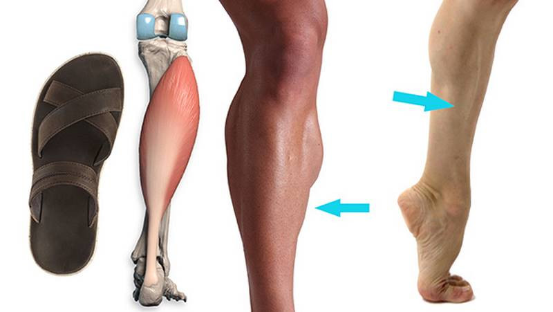
“Soleus” comes from Latin for a type of flat sandal, which is a pretty accurate description of this muscle’s form. We’ve seen “mattress” muscles like this before: brachialis in the arm, vastus intermedius in the quads. We kind of blew off vastus intermedius because it’s almost completely hidden. But the soleus is very much visible on the surface. It can be seen independent of gastrocnemius even on thin figures. It clearly pokes out from underneath the edges of gastrocnemius, especially at the ankle where the soleus muscle belly descends lower and is wider than gastrocnemius. Soleus can even be seen in front view, behind the tibia. And gastrocnemius is behind soleus. Kind of like stairsteps.
Soleus exists entirely below the knee, originating from the tibia and fibula. It’s important to notice how slanted soleus is: high on the outside, low on the inside. Gastrocnemius has this same slant. If we zoom way out, this angle fits the pattern of alternating apexes throughout the leg: the zig zag of asymmetry. These are rhythms we seek to exaggerate when we want to draw dynamic anatomy.

Roughly 2/3 down the leg, soleus begins to narrow and interlace with the Achilles tendon. It leaves a gap as it passes behind the ankles, at least one finger in width.
Gastrocnemius
Now for the superficial superstar: gastrocnemius. As important as soleus is, Mt Gastroc is more of a focal point. It’s probably the muscle you think of when you think of the calf.
The hamstring tendons create this tunnel for the gastrocnemius to squeeze into. This top region is normally hidden by a fat pad, but it’s good to know the origin anyway. Gastrocnemius originates above the knee, on the top of the femoral condyles, and it has two heads, one from each condyle. These heads tend to soften together to create a round bump. This is where the name gastrocnemius comes from: it’s “gastroc,” as in stomach, because anatomists are throwing shade and saying that the calf looks like a big round pot belly. Anyway, it really is two separate heads, and they split when the calf tenses.

These heads are not symmetrical: the lateral head ends higher and tends to create an oblique edge that flows into the Soleus. The medial head is bigger and rounder. You’ll usually see it take a sharper turn inward to attach to the Achilles tendon. In essence, the lateral head makes a slash and the medial head makes a hook. Together they kinda make an upside down heart...
The gastrocnemius is most interesting where muscle meets tendon, about halfway down the lower leg. If you’re idealizing the design, you can make the muscle longer on a female to get that sleek feminine look that artist’s often go for. And a little shorter on a male, if you’re shooting to make your model look more masculine and accent their muscularity. There’s a lot of variation in that proportion between individuals, and of course, when gastrocnemius contracts, the belly shortens so, if someone’s wearing high heels or reaching for something up high, the muscle belly will shorten.

Remember that slant: the Achilles tendon doesn’t come in like this, but at an angle. Like vastus medialis in the quads, the medial gastrocnemius head is low and heavy, and creates a round form where it meets the Achilles tendon.
Now for the calf muscle’s final form … the Achilles tendon! Despite the legends about its fragility, the Achilles tendon is actually the thickest and strongest tendon in the human body. It wraps around the lower third of the calf and funnels muscle tissue from soleus and gastrocnemius into a thick cable that anchors down on the heel bone. This creates a V shape. The narrowest point of the tendon is about 3 fingers’ width above the heel.
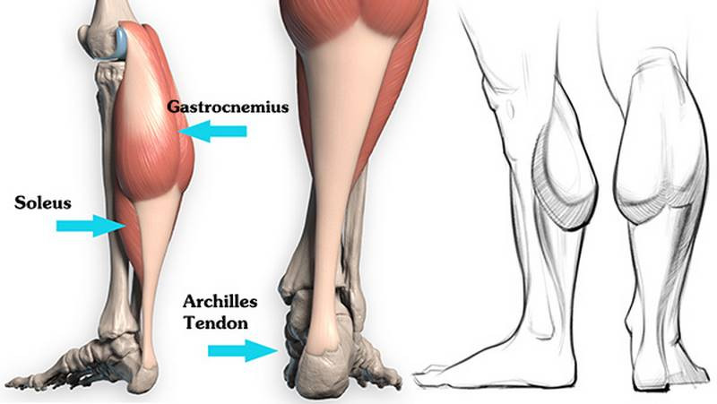
So let’s review. From inside profile, we can see how gastrocnemius is big and round, while the soleus is wide and flat. The gastrocnemius meets the Achilles tendon at around the halfway mark. If we smooth things over, the calf looks like an upside down bowling pin...or a chicken drumstick... or a caveman’s club! Except it’s slanted, high on the outside, low and heavy on the inside. When the leg tenses, it splits into three forms. The two heads of the gastrocnemius and the soleus flowing down into the Achilles tendon. And that’s what you need to know to draw the calf. Except of course all the stuff I left out of this video. That stuff will be in the premium video. In premium we’ll analyze the forms in more detail and expand on the anatomical info. And as always, you’ll get assignment demonstrations, 3d models and an ebook.
Of course, there’s only one way to really master anatomy. Get that pencil sharpened and let’s get started on your assignment.
Assignment
Your assignment is to do tonal studies of the calves from the photos provided in the description below. Start with a linear layin making sure you’re tracking everything where it should go, and then add clean tone on top to study the planar structure of the calves. Make sure to post your assignment to the community at proko.com/groups if you want to be featured in the critique video. Alright thank you guys for watching. Leave a comment below if you have any questions about the calves.


